Kyoto University has a large collection of human embryos (40,000-50,000) that were collected from 1961 to 1974. This famous "Kyoto Collection of Human Embryos" is believed to be the largest human embryo collection in the world. Because such a collection would never be obtained again, their non-destructive 3D measurements are highly desired.
MR microscopy is a very powerful tool for 3D measurements of human embryos, because chemically fixed human embryos contain large quantities of mobile or NMR visible protons, which are major components of the formalin preservation fluid. Consequently, Kyoto and Tsukuba Universities began a project in 1999 to acquire 3D MR microscopic images of thousands of embryos. We finally acquired 3D MR microscopic images of 1,204 chemically fixed human embryos using a super-parallel MR microscope operated at 2.34T.
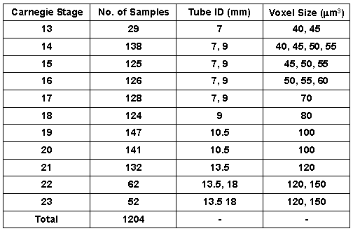
The chemically fixed human embryos measred in this project are tabulated below.