system for evaluating rheumatoid arthritis(RA), and determine its advantages
and disadvantages as an imaging modality for evaluating RA.
Materials and Methods: We prospectively studied 13 healthy controls with
no clinical symptoms of arthritis, and 13 patients with hand and wrist pains
(including pain from RA) with a 0.2T permanent-magnet compact MR imager.
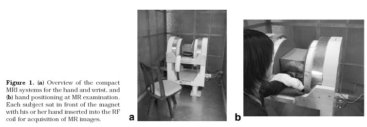
All MR images were obtained while the subjects were in a sitting position.
Coronal three-dimensional spin-echo T1-weighted images and coronal two-dimensional
short tau inversion recovery(STIR) images were obtained with image matrix=256x128
and field of view(FOV)=20.48cm.
Plain radiograph findings and MRI findings of patients were compared.
Resuits: In three of the patients with suspected early RA (N=7),
early RA was evaluated based on STIR images. All RA patients showed morphologic
or signal intensity changes that allowed an evaluation of RA from MR findings.
Four of five RA patients showed high signal intensity on STIR images in the wrist,
proximal interphalangeal(PIP) joint, or metacarpophalangeal(MCP) joint that
suggested synovitis. Multiple erosions in the hand and wrist were seen in four RA patients,
with low signal intensity on T1-weighted images.
Conclusion: RA was correctly evaluated, and early RA could be identified with
the compact MRI system. However, the current system has limitations,
such as the nonselective STIR sequence used and magnetic field inhomogeneity.