MR Fingerprinting and Applications
MRI is indispensable in the medical field, but those who have undergone MRI examinations may have the impression that it is time-consuming; MRI examinations generally take about 30 minutes, and this is due to the fact that multiple methods of imaging are performed separately.
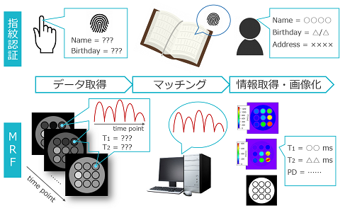
MRI can measure various tissue parameters, such as nuclear magnetization relaxation times (T1 and T2), proton density, and diffusion. In the past, these parameters were generally measured by different imaging methods. A new imaging method called MR Fingerprinting (MRF) was published in Nature in 2013. The reason for the name "Fingerprinting" is that the sequence of steps from imaging to matching and quantitative mapping can be likened to the process of fingerprint identification.
In our laboratory, we are implementing MRF in our equipment in our laboratory, and are also conducting experiments to apply it to bone and soft tissue. Furthermore, we are devising methods of signal acquisition and image reconstruction using simulations, aiming to further increase the speed of MRF while employing a simple data acquisition method that can be implemented in clinical MRI systems. If applied to medical practice, MRF is expected not only to reduce the burden on patients, but also to significantly shift the paradigm of diagnostic imaging from conventional qualitative evaluation to quantitative evaluation.
担当・文責:佐々木 椋一 (2018年4月12日)